
How Much Is an Urgent Care Vet Visit? Costs and What to Expect
The cost of an urgent care vet visit can vary depending on your pet’s condition, the care they need, and the clinic you visit. While many pet owners look for a simple number, the reality is that urgent care pricing is based on the level of care provided during the visit. Some visits are quick […]
Vet Urgent Care vs Emergency Vet | What’s the Difference?
When your pet is sick or injured, one of the hardest decisions is figuring out where to go. Should you head to urgent care or go straight to an emergency vet? The answer depends on how serious the situation is. While both provide important care, they are designed for very different levels of need. Knowing […]
When to Worry About Puppy Diarrhea | Warning Signs to Know

Puppy diarrhea is common, and in many cases, it is mild and short lived. Puppies have sensitive digestive systems, and things like diet changes, stress, or eating something unusual can easily cause loose stool. If your puppy is still eating, drinking, and acting normal, the issue may resolve on its own within a day or […]
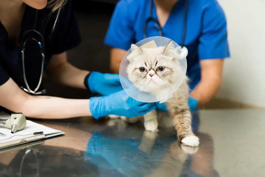
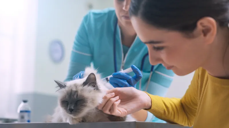
How to Treat a Cat Skin Infection | Symptoms and Care

Cats are great at hiding discomfort, which means skin infections can go unnoticed until they become more advanced. What may start as mild irritation can quickly turn into something more serious if left untreated. Understanding what to look for and how to respond early can help your cat heal faster and stay comfortable. What a […]
Why Is My Dog Limping on Its Back Leg?

One minute your dog is running around like normal, and the next they are limping on their back leg. It can be alarming, especially if you are not sure what caused it. Limping is a sign that something is uncomfortable, painful, or not working properly. Sometimes it is minor and temporary, but other times it […]
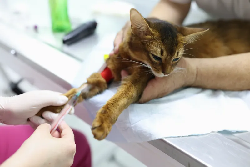
Does Pet Insurance Cover Urgent Care? What to Know
Most pet insurance plans do cover urgent care visits, but it depends on your specific policy. Coverage typically applies if the visit is related to an accident or illness that is not pre-existing. However, what gets reimbursed and how much you get back can vary based on your plan details. Understanding how your coverage works […]
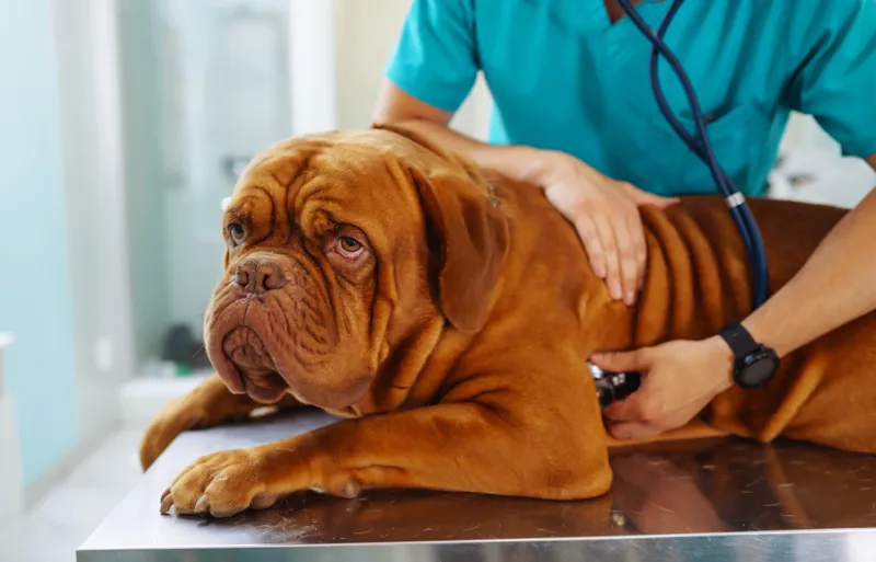
Signs Your Dog is Sick | When to Worry and What to Do
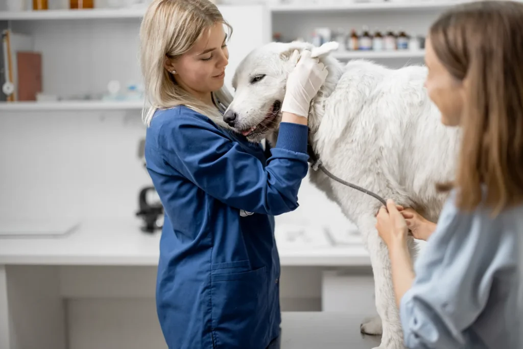
Dogs cannot tell you when something feels wrong, which is why it is so important to notice small changes. In many cases, the early signs of illness are subtle and easy to miss. A dog that seems slightly “off” today could be showing the first signs of a bigger issue. Paying attention early can make […]
Cat Throwing Up? When to Take Your Cat to the Vet
Cats throw up more often than many pet owners expect. Sometimes it is as simple as a hairball or eating too quickly, and your cat goes right back to normal like nothing happened. But vomiting can also be one of the earliest signs that something more serious is going on. The challenge is knowing when […]
Puppy Has Diarrhea But Still Eating and Drinking? | What to Do

Puppy has diarrhea, but is still eating and drinking If your puppy has diarrhea but is still eating and drinking, it can feel confusing. The good news is that this is often a sign the issue may be mild, especially if your puppy is still active and alert. That said, diarrhea is never something to […]
Do Dogs Need Pain Meds After Neuter Surgery?

Many pet owners wonder whether dogs need pain meds after neuter surgery. Neutering dogs is one of the most common veterinary procedures, and while the surgery is routine, dogs can still experience pain as their bodies heal. Most veterinarians provide pain medications as part of the recovery process to manage discomfort, keep your dog comfortable, […]